




















































Advanced digital imaging is transforming dental surgery, offering greater precision and helping to make procedures safer for patients. Modern scans and imaging technologies are now fundamental in diagnosis, treatment planning, and patient communication, providing higher-quality data and streamlining clinical workflows. These developments are shaping contemporary dental care practices.
In dental practices, the integration of digital imaging has changed both clinical methods and patient experiences. Compared with traditional dental techniques, today’s digital tools—such as intraoral scanners, cone beam computed tomography, and digital photography—provide enhanced accuracy, comfort, and efficiency. In the context of oral surgery on Harley Street, these innovations support clinicians by offering critical information for complex diagnoses and treatments. As dental professionals continue to implement digital technology, patients benefit from clearer diagnosis, more predictable planning, and precise treatment delivery across a variety of oral surgery procedures.


Digital imaging technologies used in dental practice
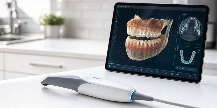
Digital imaging includes a range of specialised tools now widely used in dental surgeries. Intraoral scanners have largely replaced physical impressions, capturing detailed 3D maps of teeth and soft tissue without the discomfort of traditional moulds.
Cone beam computed tomography (CBCT) provides three-dimensional cross-sectional images of the jaw, sinuses, and supporting structures. These scans deliver much more detail than standard 2D X-rays, improving the quality of surgical planning and diagnostics in dental and oral surgery.
Digital photography, taken both inside and outside the mouth, documents tooth surfaces, gum condition, and facial features. This supports clear case documentation and effective communication among dental professionals.
How digital imaging aids diagnosis and treatment
The use of digital imaging in dental settings improves early detection of conditions such as decay, cysts, or bone loss. High-resolution images enable clinicians to identify subtle anatomical changes that might be missed with standard films or clinical exams.
Digital scans enhance visualisation of complex anatomical features, which is essential in procedures involving nerves, sinuses, or impacted teeth. This information supports the creation of accurate, individualised treatment plans and helps to anticipate potential complications, reducing risk in surgery.
For patients requiring specialist care, including those who may see an emergency dentist in Liverpool, digital imaging delivers the necessary data for timely assessment and intervention. This allows dental teams to make informed, confident decisions when urgent treatment is required.
The ability to access and compare past and current digital records allows clinicians to monitor changes over time with greater accuracy, supporting ongoing care throughout every stage of treatment.
Improving workflows and patient experience in surgery
Digital imaging works alongside computer-aided design and virtual planning systems. These tools allow for step-by-step simulation of surgical procedures, so clinicians can plan thoroughly and coordinate their teams using clear visual references before starting treatment.
For patients, digital scans often provide greater comfort by removing the need for impression materials. Many individuals find intraoral scanning less invasive, resulting in faster appointments and reduced anxiety, particularly for those with sensitive gag reflexes or dental anxiety.
Digital imaging also supports better patient communication. Visual models and scans help patients understand their condition, treatment options, and the expected results of oral surgery, enabling more informed consent and setting realistic expectations.
While digital imaging is suitable for many procedures, traditional methods remain necessary in certain situations. Some anatomical or technical factors may require conventional impressions or radiographs, so maintaining a balanced approach ensures the best clinical outcomes.
Data security, governance, and looking ahead
Digital records offer reliable documentation for both clinicians and patients. These images can be securely stored, retrieved quickly, and shared electronically as needed to support ongoing care and collaboration between specialists.
Dental surgery providers must manage data responsibly, maintaining secure storage and complying with legal standards for data protection to safeguard patient privacy.
In the future, artificial intelligence may support dental imaging by assisting with scan interpretation and diagnostic work. Ongoing development of integrated software and planning tools could further improve clinical workflows, though any advances require careful review and appropriate regulation before wider adoption.
Digital imaging has become a vital element of modern dental surgery, continuing to evolve in support of better patient care, safety, and responsible management of clinical data.

David Prior
David Prior is the editor of Today News, responsible for the overall editorial strategy. He is an NCTJ-qualified journalist with over 20 years’ experience, and is also editor of the award-winning hyperlocal news title Altrincham Today. His LinkedIn profile is here.